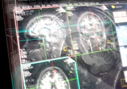
| A team of Korean scientists said they have developed a technology to use nano-particles to draw clinical images of cells involved in cancer, which could bring a breakthrough in the early detection of the disease. Researchers led by Cheon Jin-woo and Seo Jin-seok said in a paper published on the Internet site of Nature Medicine on Monday that their new technology allowed magnetic resonance imaging (MRI) scans to find cancer cells less than 2 millimeters in size. Conventional MRI tests could only distinguish cells of a larger size, which are found only after the cancer is further developed. A nano-particle is a microscopic particle whose size is measured in nanometers, or one billionth of a meter. Recent scientific research has proved nano-particles to be useful in detecting tumors due to their abilities to act as imaging agents and make cells and tissues more visible in MRI scans. The nano-particles developed by the Korean researchers could be swallowed like a pill or injected through a catheter into the human body. Their function is to detect cancer and other diseases in their early developmental stages. Among the commonly used nano-particles in MRI imaging is CLIO, which was developed by scientists at Harvard University. In their paper in Nature Medicine, the Korean scientists claimed that their new nano-device performed better than CLIO in MRI experiments, generating a signal that was about 10 times more intense.
|


